bret
Big Boned
Published: January 31st 2023, 11:48:53 pm
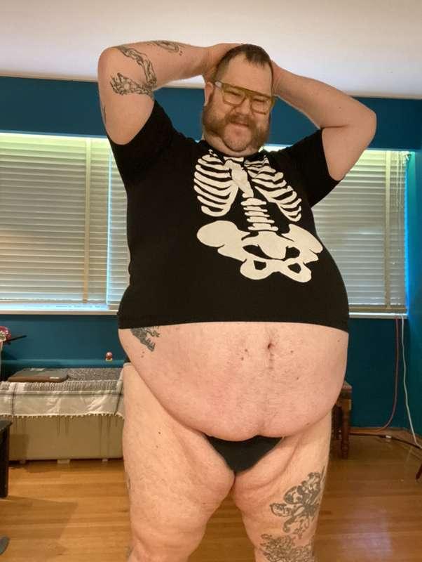
I did some imaging recently of my knees and ribs for some fat boy trubs. I’m an xray tech so I personally find this stuff fascinating. Thought you all may be interested to see what very fat knee and chest xrays look like. 😉
P.S. I’m fine.
1st image is a front view (Antero-Posterior, AP) of my knee.
2nd is a sideways view (lateral) of my knee.
3rd image is a view shot up through my knee cap (skyline/retropatellar).
4th image is a PA view of my chest.
5th is a lateral of my chest.
I love seeing how different my body is to a normal size person. The chest xrays have almost no black space around them because my fat body fills in all that extra space.
Xrays images are essentially measures of 3D density compressed into a 2D image. The whiter an area is the more density exists in the body in that area. Bones are very dense; air-filled lungs, not so much. Unfortunately for obese people, in order to get a good quality image we need to apply more xrays to get through all the thickness we have. My chest xrays needed about 3 times as much radiation for my PA chest and about 6 times as much for my lateral chest than a normal size person would need. Although, doses these days are pretty low compared to what they used to be. More efficient processing algorithms reduce necessary dose quite a lot, so it’s not that bad.
That being said, I did also do a seperate specialized rib view that goes through a lot of my belly though (not pictured). Since I’m so fat it maxed out the exposure possible in one of our rooms. I think we hit 160mAs. This measurement is a value that is proportional to the quantity of xrays. My knee xray took 6mAs for reference.
Anyway, enough nerding out.
If you have any questions about medical imaging, I’d love to answer them. My pictures, or the process, or your images if you have them.
Let me know.
Love you, Byeeee